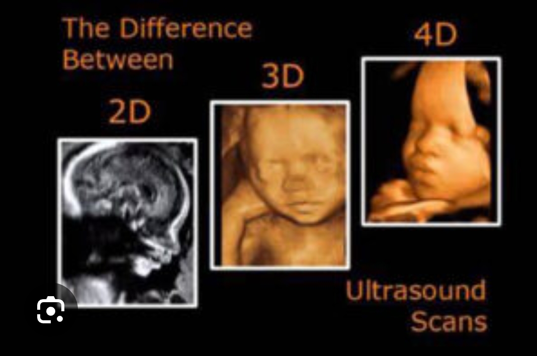
3D and 4D ultrasound scans are advanced imaging techniques used during pregnancy to give more detailed views of the baby compared to the standard 2D scan.
Here’s a simple breakdown:
🔹 What is a 3D Scan?
A 3D ultrasound creates a three-dimensional image of your baby. Instead of a flat black-and-white image (like a regular 2D scan), it shows depth — similar to a photograph.
🩺 Role of 3D Scan in Pregnancy:
Detects structural abnormalities
Cleft lip and palate
Facial abnormalities
Limb defects
Spine problems
Better visualization of organs
Brain
Heart
Kidneys
Helps doctors explain findings to parents
Easier to understand than 2D images
Bonding experience
Parents can see baby’s face clearly
🔹 What is a 4D Scan?
A 4D ultrasound is basically a 3D scan in motion — it shows real-time movement.
You can see:
Baby smiling
Yawning
Stretching
Moving hands and legs
🩺 Role of 4D Scan in Pregnancy:
Assess fetal movements
Helps evaluate muscle tone and neurological development
Detect facial and limb abnormalities more clearly
Evaluate heart movement and function (in some cases)
Provides reassurance to parents
📅 When Are They Done?
Usually between 26–32 weeks for best facial images
Can be done earlier if there is a medical indication
⚠️ Are They Necessary?
They are not routine for all pregnancies.
Standard 2D ultrasound is sufficient for most medical evaluations.
3D/4D scans are mainly used when:
A structural abnormality is suspected
High-risk pregnancy
Detailed assessment is needed
✅ Are They Safe?
Yes — they use the same ultrasound technology (sound waves, not radiation) as 2D scans and are considered safe when performed by trained professionals.