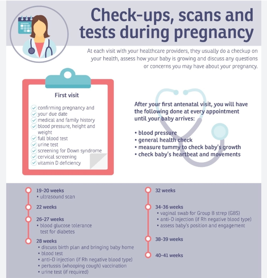
Here’s a list of the important ultrasound scans and imaging typically done during pregnancy, along with their timing and purpose:
📅 Key Pregnancy Scans & Imaging Tests
1. 🧫 Dating Scan (6–9 weeks)
Purpose: Confirm pregnancy, estimate gestational age, check for heartbeat and number of fetuses.
Often done via transvaginal ultrasound if early.
2. 📏 Nuchal Translucency (NT) Scan / First Trimester Screening (11–13+6 weeks)
Purpose:
Measure fluid at the back of the baby’s neck (nuchal fold).
Combined with blood tests to assess risk of Down syndrome (trisomy 21) and other chromosomal conditions.
Often part of the combined screening.
3. 🧠 Anomaly Scan / Mid-Trimester Morphology Scan (18–22 weeks)
Purpose:
Detailed structural assessment of the baby’s organs and limbs.
Check the position of the placenta, amniotic fluid, and fetal growth.
Rule out congenital anomalies (heart, brain, spine, kidneys, etc.).
4. 📊 Growth Scan(s) (28–32 weeks or later if needed)
Purpose:
Monitor fetal growth, weight, and development.
Assess amniotic fluid levels and placental function.
Common in high-risk pregnancies (e.g., gestational diabetes, hypertension, IUGR).
5. 🧪 Doppler Ultrasound (If indicated, often around 28–36 weeks)
Purpose:
Evaluate blood flow in the umbilical cord, fetal brain, or placenta.
Used in cases of suspected fetal growth restriction, preeclampsia, or other complications.
Optional / Additional Scans:
🧬 Chorionic Villus Sampling (CVS) (11–13 weeks)
For genetic diagnosis in high-risk pregnancies.
🧬 Amniocentesis (15–20 weeks)
Diagnostic test for genetic/chromosomal abnormalities.
💓 Fetal Echocardiography (18–24 weeks)
Specialized scan of the fetal heart, often done if there's a family history or abnormality found in anomaly scan.
🧍♀️ Cervical Length Scan (16–24 weeks)
For women at risk of preterm labor