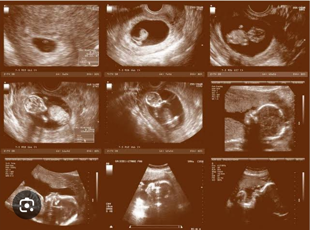
During pregnancy, ultrasounds (sonograms) are used to monitor the development of the baby and the health of the mother. There are typically three main ultrasound scans, one for each trimester, though some women may have more depending on their health or circumstances.
1. First Trimester (Weeks 1–12)
Purpose: This ultrasound is usually performed between 6-12 weeks to confirm the pregnancy, verify the due date, detect the number of embryos (singleton or multiples), check the heartbeat, and evaluate the position of the embryo.
Types:
Transvaginal Ultrasound: Often used early in pregnancy for a clearer view of the embryo.
Abdominal Ultrasound: Used later in the first trimester to confirm findings from earlier scans.
2. Second Trimester (Weeks 13–26)
Purpose: The mid-pregnancy ultrasound (often called the "anatomy scan") is performed between 18-22 weeks. It checks the baby's growth, the development of organs and structures, and verifies the position of the placenta.
Types:
Abdominal Ultrasound: Most common in this trimester, performed on the abdomen using gel and a probe to create images of the baby.
Gender Reveal: This ultrasound may also reveal the baby's gender, although it's not the primary purpose.
3. Third Trimester (Weeks 27–40)
Purpose: Ultrasounds in the third trimester are typically performed if there are concerns about fetal growth, amniotic fluid levels, or the baby's position before delivery. It may also be used to check for any complications such as placental issues.
Types:
Abdominal Ultrasound: Most common for monitoring fetal growth and position.
Doppler Ultrasound: Sometimes used to assess blood flow in the umbilical cord and check the baby's health.
While these are the typical scans during each trimester, additional ultrasounds may be performed if the pregnancy is high-risk or if there are complications.
Keywords
1822 weeks
612 weeks
circumstances 1
typically performed
trimester performed
babys health
multiples check
confirm findings
babys position
pregnancy verify
umbilical cord
primary purpose 3
create images
anatomy scan
clearer view
embryos singleton
midpregnancy ultrasound
typical scans
earlier scans 2
babys gender
babys growth
baby gender reveal
position doppler ultrasound
main ultrasound scans
trimester additional ultrasounds
pregnancy ultrasounds sonograms
embryo abdominal ultrasound
monitoring fetal growth
assess blood flow
due date detect